Past Retreats: 2014 Image Competition Entries
Click to view larger images.
The Committee also considered Monica Gupta's submissions from 2013 and Luan Wen's submissions from 2012.
Competition Winner:
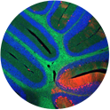
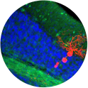
Ian Williams, PhD
Section on Molecular Dysmorphology (Forbes Porter Lab)
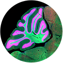
Degeneration in cerebellum: Brain sections from our NPC1 mouse model. Blue = DAPI, Green = lipid stain, Red = Neuron Stain.
Monica Gupta, PhD
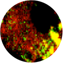
Section on Molecular Genetics of Immunity (Keiko Ozato Lab)
The image is of bone marrow derived macrophage immunostained with MDC (green, autophagy marker) and mitochondrial dye (red) respectively. The overlapping image shows the presence of depolarized/damaged mitochondria in autophagosomal compartments of the cells that are eventually degraded by the process of autophagy.
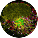
Alex Valm, PhD & Sarah Cohen, PhD
Section on Organelle Biology (Jennifer Lippincott-Schwartz Lab)
Six-color image of peroxisomes (cyan), Golgi (green), endoplasmic reticulum (yellow), mitochondria (red), lysosomes (magenta), and lipid droplets (white) in a live Cos7 kidney cell.
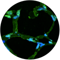
Ping Wang, PhD (William J. Martin II Lab)
Unit on Lung Injury and Repair
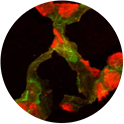
Frozen section of murine lung with immunofluorescent stain with SPC (in green) for identification of type II alveolar epithelial progenitor cells (AT II) on day 2 and 14, respectively, following intratracheal instillation of bleomycin. Note the shape of SPC(+) AT II cells changed from cuboidal on day 2 to flat shapes on day 14 after bleomycin, indicating that AT II cells replaced flat-shape ATI cells in alveoli during lung repair after bleomycin-induced lung injury.
Ping Wang, PhD (William J. Martin II Lab)
Unit on Lung Injury and Repair
Confocal image of frozen section of murine lung on day 21 following intratracheal instillation of bleomycin and airway delivery of donor DiI-labeled alveolar epithelial progenitor cells (AEPCs) (in red) on day 4 after bleomycin. Note the DiI-labeled donor AEPCs revealed in alveolar wall, indicating donor AEPC engraftment with the alveolar cells in the lung of the recipient mouse after airway delivery.
Ping Wang, PhD (William J. Martin II Lab)
Unit on Lung Injury and Repair
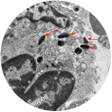
Transmission electron microscopic (TEM) image of murine lung on day 11 following intratracheal instillation of bleomycin. It revealed an eosinophil degranulation (red arrows) during the eosinophil penetration of arteriolar endothelium.
Ian Williams, PhD
Section on Molecular Dysmorphology (Forbes Porter Lab)
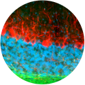
Brain sections from our NPC1 mouse model. Blue = DAPI, Green = lipid stain, Red = Neuron Stain.
Ian Williams, PhD
Section on Molecular Dysmorphology (Forbes Porter Lab)
Brain sections from our NPC1 mouse model. Blue = DAPI, Green = lipid stain, Red = Neuron Stain.
