
Past Retreats: 2012 Image Competition Entries
Click to view larger images.

Competition Winner:
Takaaki Miyazaki, PhD
Section on Sensory Coding and Neural Ensembles (Mark Stopfer Lab)
Images show three views of an olfactory projection neuron in the brain of an adult locust: Dorsal (top), anterior (down left) and lateral (down right). The anterior view shows three groups of neural fibers: the neuron's output areas in the mushroom body calyx (top left) and the lateral horn (top right), and the input area, the antennal lobe (bottom), which contains the glomeruli, which are spherical neuropil found in both vertebrate and invertebrate olfactory systems. The neuron was labeled with neurobiotin injected through a glass microelectrode and was visualized with avidin-conjugated Alexa 633 fluorophore. Confocal data acquired with Zeiss LSM 510 Inverted Meta of NICHD-MIC were processed with ImageJ and FluoRender (ver. 2.9.1) software.
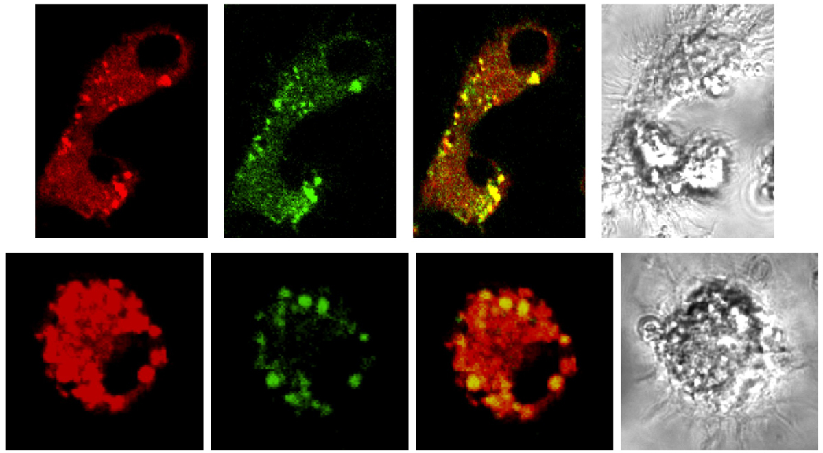
Monica Gupta, PhD
Section on Molecular Genetics of Immunity (Keiko Ozato Lab)
These are images of Bone marrow derived Macrophages stained with Dye MDC (Green) specific to detect Autophagosomal/lysosomal compartments and Mitochondrial Dye (MT Deep Red). The overlapping image shows the presence of depolarized/damaged Mitochondria to be degraded by the cell in especial pin pointed structures called Autophagosomes.
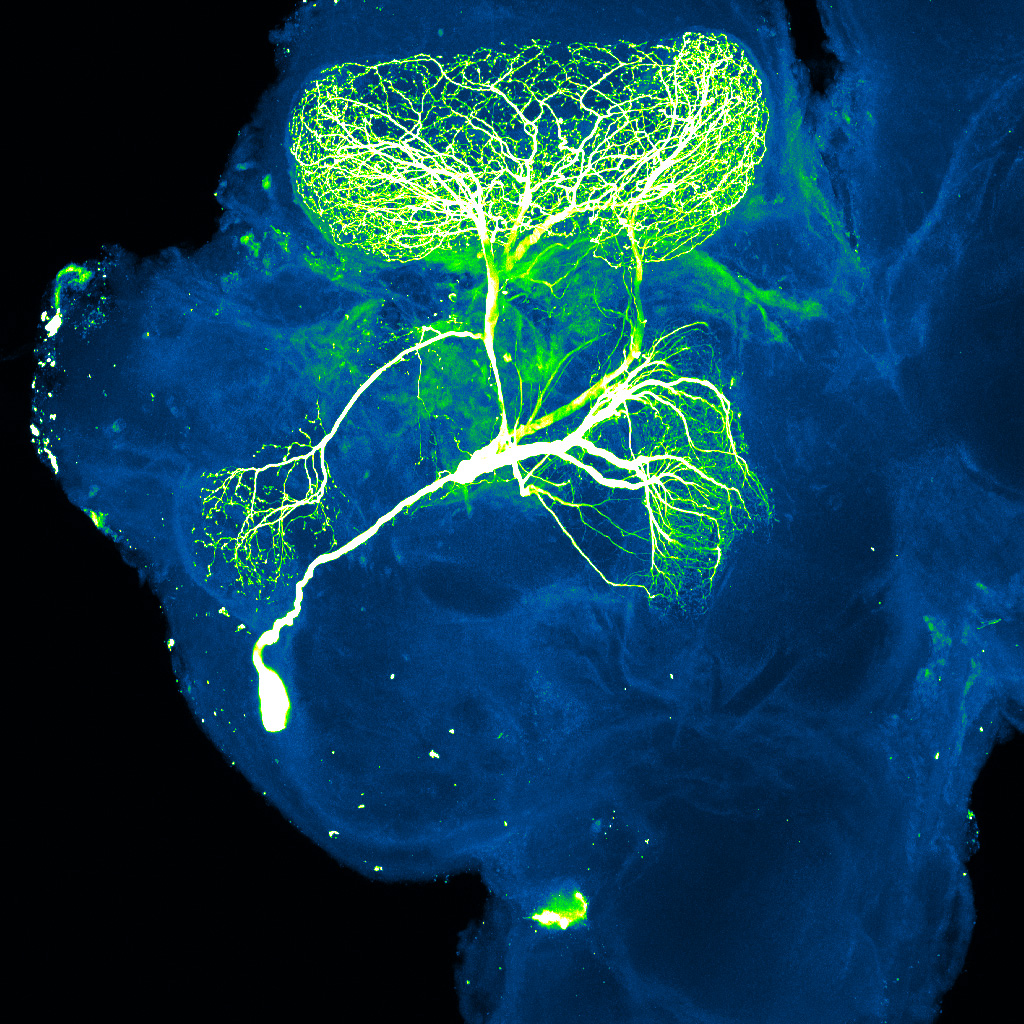
Nitin Gupta, PhD
Section on Sensory Coding and Neural Ensembles (Mark Stopfer Lab)
Image shows a neuron (called 'Giant GABAergic Neuron') in green/white, in one-half of an insect brain (blue).
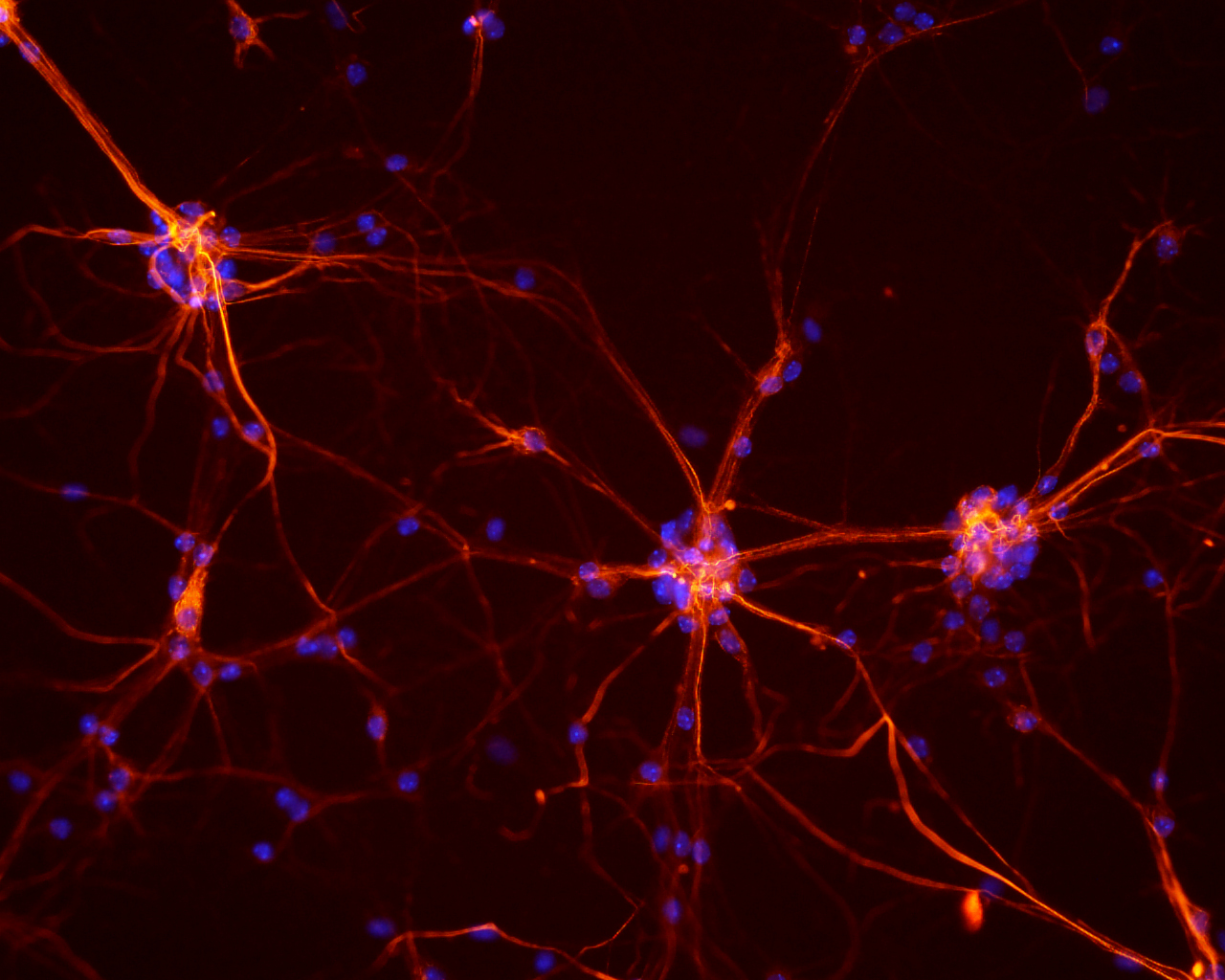
Suh-Young Jeong, PhD
Section on Human Iron Metabolism (Tracey Rouault Lab)
Neurons from mouse spinal cord primary culture were stained with a cocktail of anti-neurofilament antibodies (SMI 310 & 32, Covance) and then visualized using Alexa Fluor 546-conguated secondary antibodies. Blue color represents nuclei stained with DAPI.
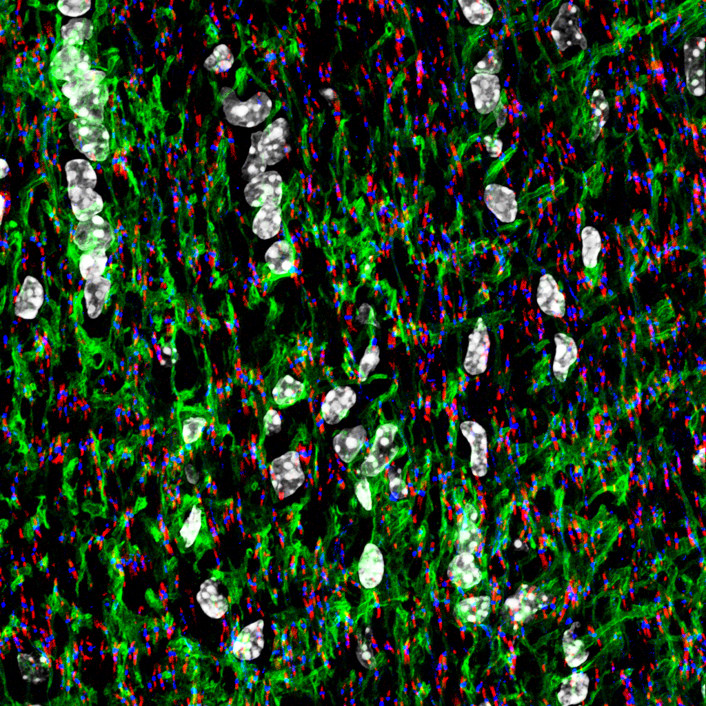
Hae Ung Lee, PhD
Section on Nervous System Development and Plasticity (Douglas Fields Lab)
"Nodes of Ranvier in optic nerve." Green is GFP expressing Astrocytes. Red is Caspr; marker for paranodes. Blue is Sodium channel; marker for nodes. White is Nucleus.
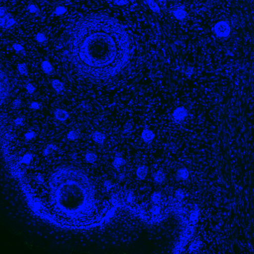
Margaret Ochocinska, PhD
Section on Neuroendocrinology (David Klein Lab)
"Starry Night"
The image is a DAPI (blue) stained mouse brain section through the olfactory bulb.
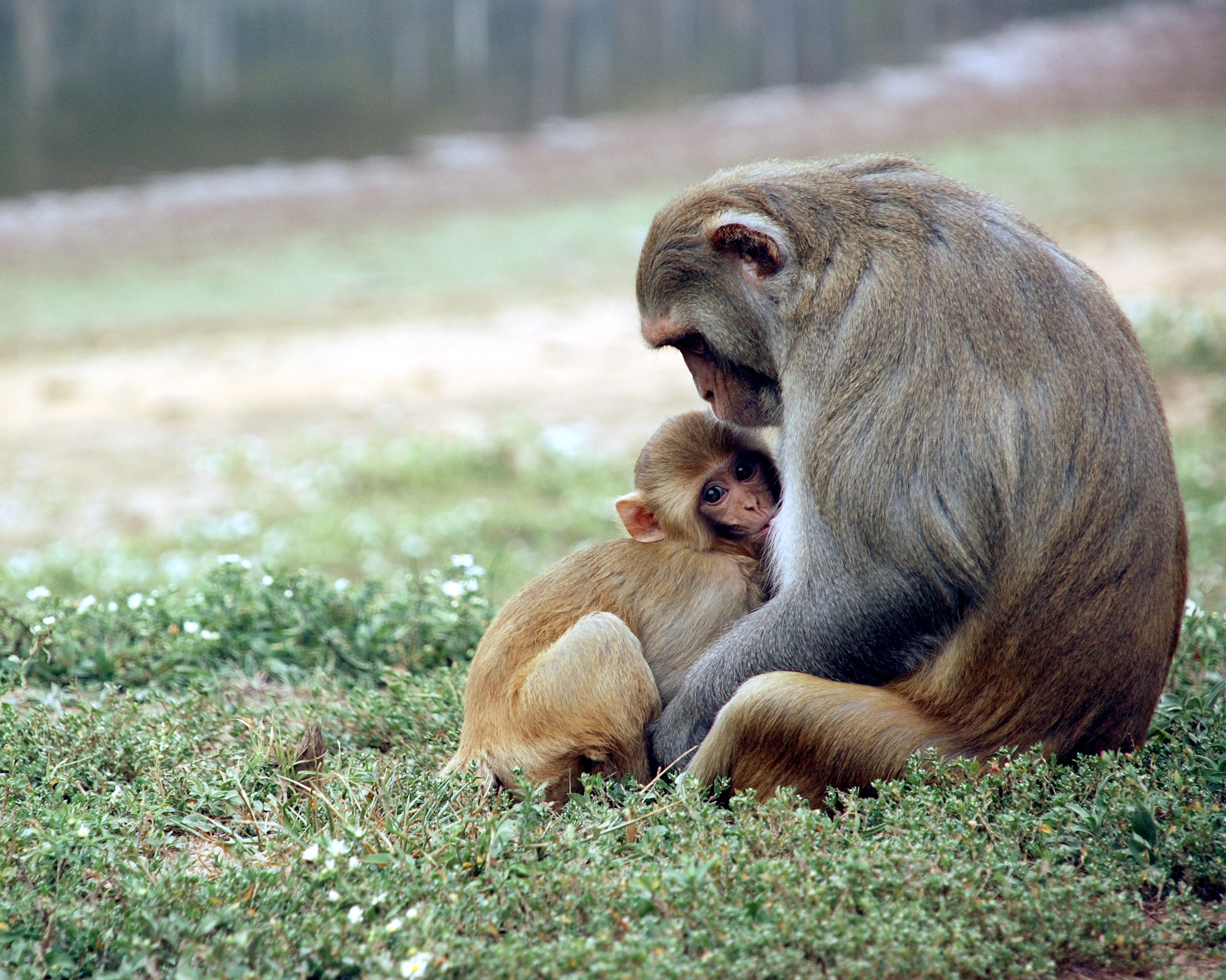
Annika Paukner, PhD
Laboratory of Comparative Ethology (Steve Suomi Lab)
"A rhesus macaque mother, Heather, nurses her 6-month-old infant, Kayla, in the field station enclosure at the NIH Animal Center, Poolesville."
Photo credit: Michelle Miller

Jason Riley, PhD
Section on Analytical and Functional Biophotonics (Amir Gandjbakhche Lab)
"How your brain thinks in flatland: is an image about how we look at things, it represents the surface of the brain color coded by 'functional' regions given by the Talairach atlas."
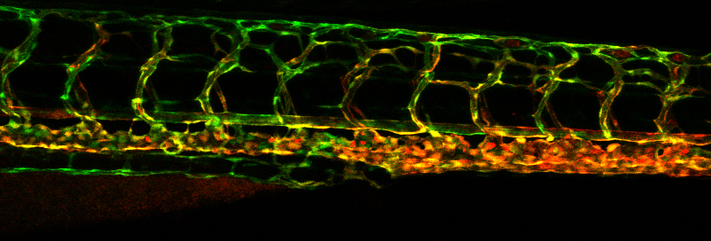
Matthew Swift, PhD
Section on Vertebrate Organogenesis (Brant Weinstein Lab)
Two color confocal image showing the lateral view of a 5 day old Tg(fli1a:eGFP);Tg(kdrl:mCherry) transgenic zebrafish trunk displaying a spontaneous mutation that resulted in overbranching of the dorsal portion of the Intersomitic Vessels (ISVs).
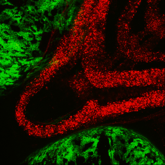
Valerie Virta, PhD
Section on Vertebrate Development (Tom Sargent Lab)
This is a ventral view of the zebrafish jaw using a fluorescent red reporter line for craniofacial neural crest cells. The eyes have been false-colored green although they are autofluorescent at this wavelength.
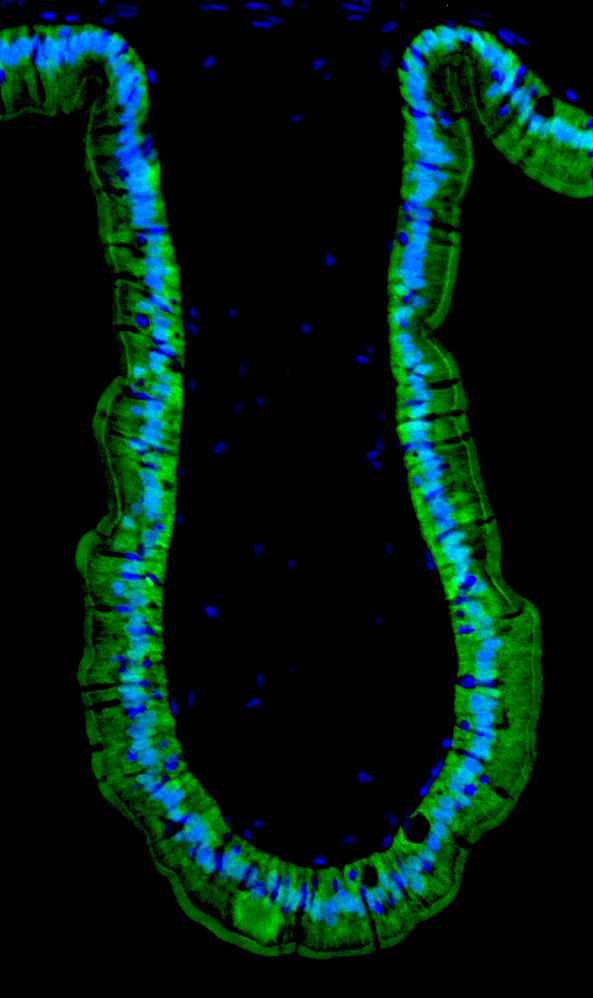
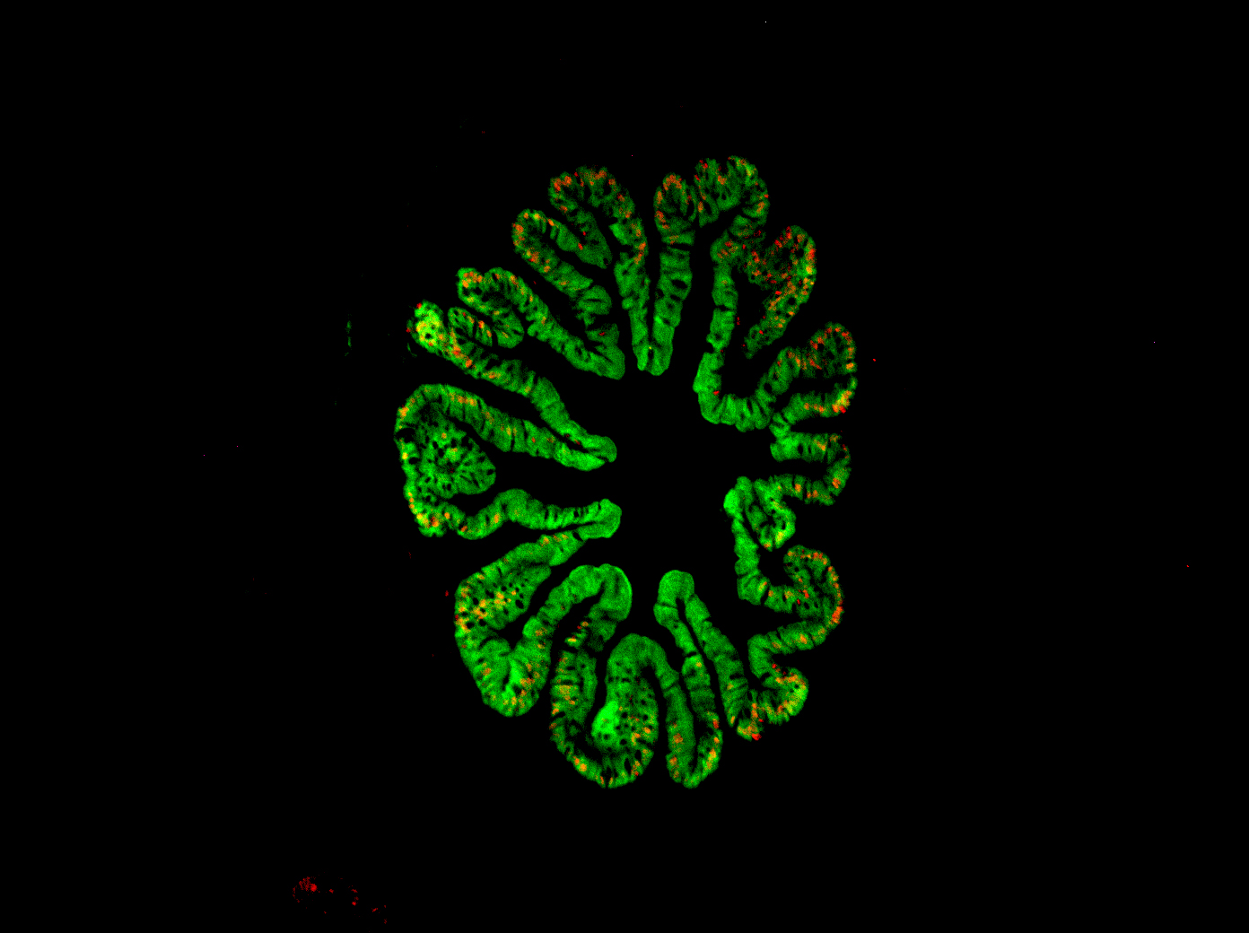
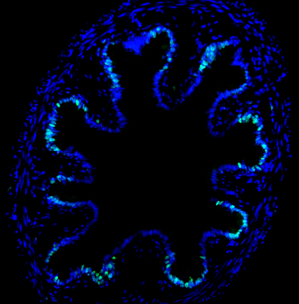
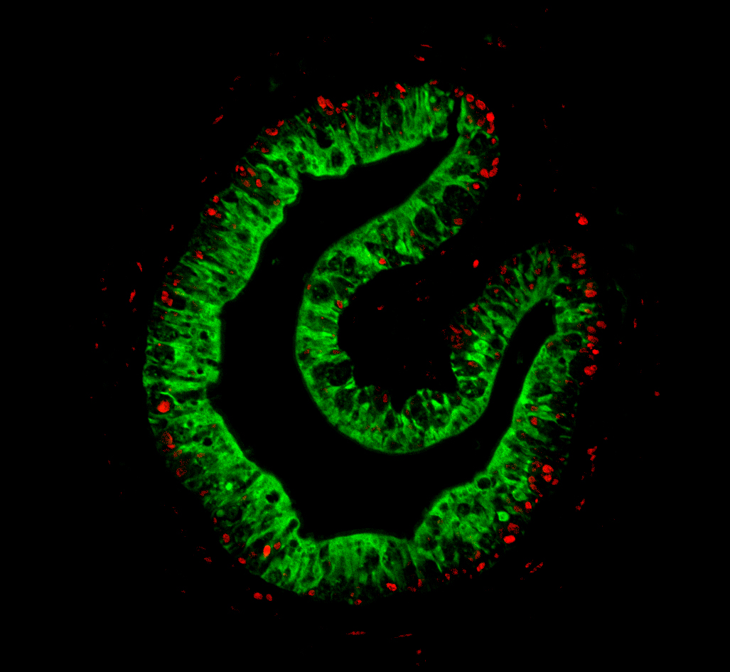
Luan Wen, PhD
Section on Molecular Morphogenesis (Yun-Bo Shi Lab)
Luan Wen, PhD
Section on Molecular Morphogenesis (Yun-Bo Shi Lab)
Luan Wen, PhD
Section on Molecular Morphogenesis (Yun-Bo Shi Lab)
Luan Wen, PhD
Section on Molecular Morphogenesis (Yun-Bo Shi Lab)