
Past Retreats: 2018 Image Competition Entries
Click to view larger images.
The Committee also considered 2017 submission from Hyun Min Jung.
Competition Winner:
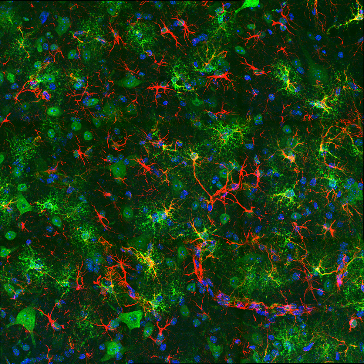
Jacob Gluski, BA
Unit on the Development of Neurodegeneration (Le Pichon Lab)
A Glia’s Dance: An ex vivo slice of spinal cord from a mouse model of neurodegeneration. Our lab is looking at the development of neurodegeneration. This is an early stage of the model. Dapi is blue, TDP-43 is green, and GFAP is red.
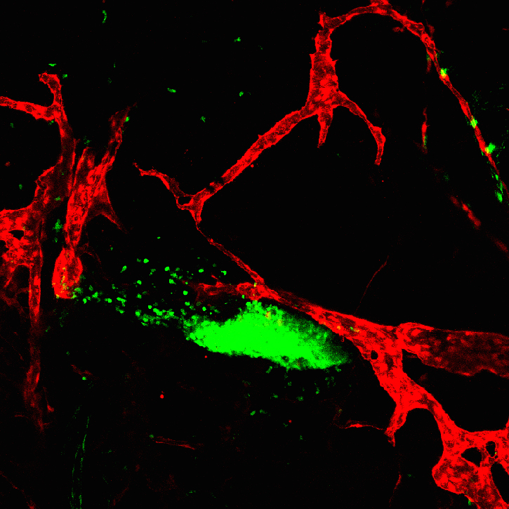
Hyun Min Jung, PhD
Section on Vertebrate Organogenesis (Weinstein Lab)
Resubmission from 2017.
Migration of lymphocytes from thymus (GFP labeled oval shape structure) toward lymphatic vessels (labeled with RFP). Lck promoter drives GFP expression and lyve1 promoter drives RFP expression.
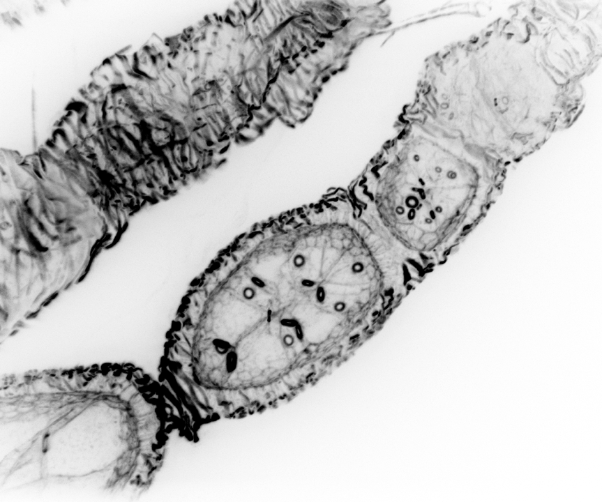
Karen Plevock Haase, PhD
Section on Cell Cycle Regulation (Dasso Lab)
Phalloidin labels actin structures in the Drosophila ovary. Germ cells undergo incomplete cytokinesis and retain an actin ring between cells.
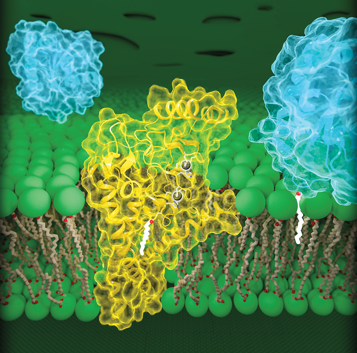
Mitra Rana, PhD
Unit on Structural and Chemical Biology of Membrane Proteins (Banerjee Lab)
Molecular view of DHHC palmitoyltransferases. Human DHHC20 palmitoyltransferase (yellow) shown localized in the Golgi body membrane (green stacks). The Zn2+ ions are shown as gray spheres and the acyl chain of the palmitoyl group in white sticks. A hypothetical substrate (blue) approaches the palmitoyltransferase from the left and, after palmitoylation, is localized to the Golgi body membrane through anchoring of the palmitoyl group, now transferred to the substrate.
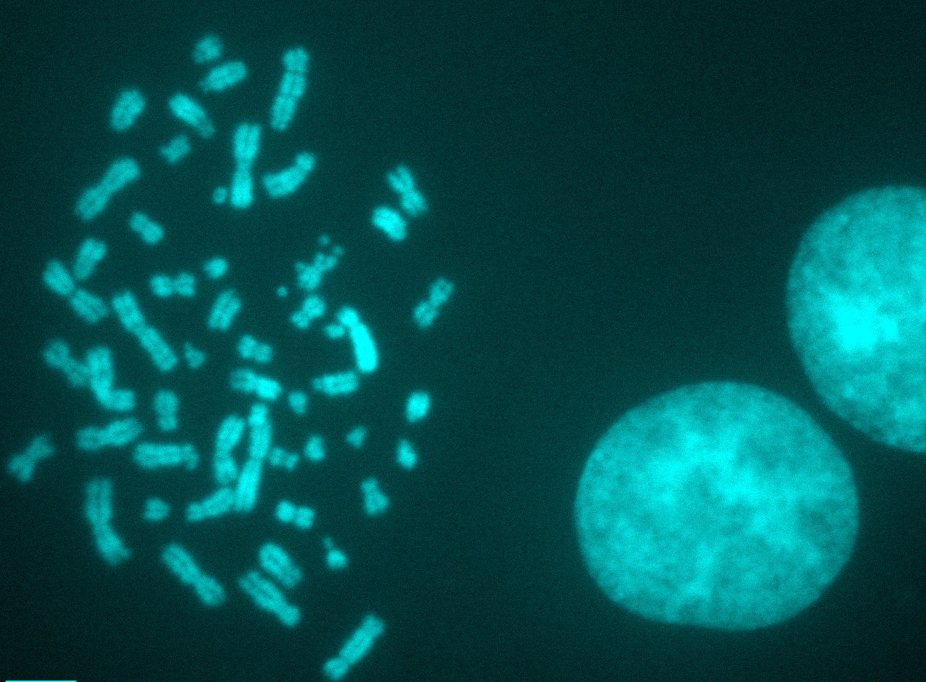
Saroj Regmi, PhD
Section on Cell Cycle Regulation (Dasso Lab)
Chromosomes: Chromosomal spread of U2OS cells showing various chromosomes on the left and intact nuclei on the right.

Saroj Regmi, PhD
Section on Cell Cycle Regulation (Dasso Lab)
Bouquet of flowers: Immunostaining of human colorectal cancer cells (DLD cells) with DNA visualized in red and unspecific antibody in green.