
Past Retreats: 2017 Image Competition Entries
Click to view larger images.
The Committee also considered 2016 submissions from Edra London and Qi Wang.
Competition Winner:
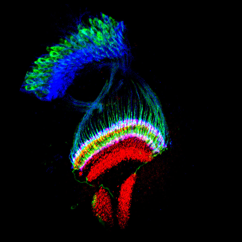
Jiangnan Luo, PhD
Section on Neuronal Connectivity (Lee Lab)
Thin section of the optic lobe of a pupal Drosophila brain (40 hours after puparium formation). Axons of photoreceptors (Blue) and lamina neurons (Green) bypass the first neuropil - lamina and project into different layers of the second neuropil - medulla (Red, center), where the visual information is integrated and processed and further relayed to higher order neuropils - lobula complex (Red, under medulla). The medulla neuropil is well organized in columnar and stratified structure, typically seen in the human visual system.
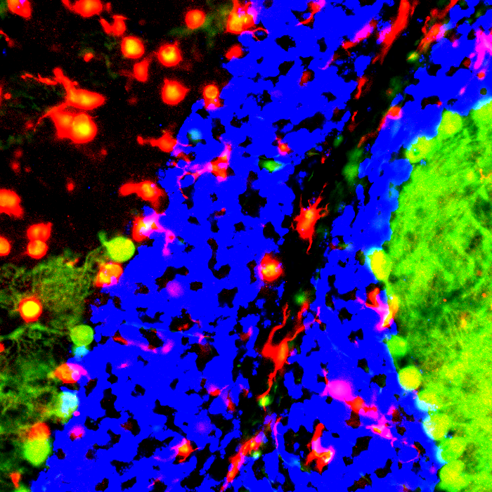
Antony Cougnoux, PhD
Section on Molecular Dysmorphology (Porter Lab)
Microglia activation in Niemann-Pick type C1 cerebellum.
This is a four color image. Green is for calbinin which in this picture stain neurons called purkinje cells, the red is for iba1 a microglia marker, the yellow is for cd68 a lysosome and activated immune cell marker and in blue is a pan nuclear staining overexposed here to make a border between two areas. On the left site of the image a highly inflammatory area with less purkinje cell and on the right a lower inflammation associated with increased number of neurons.
This picture is from the cerebellum of a mouse recapitulating the Niemann-Pick type C1 disease known to have neuroinflamamtion in specific area of the brain. The role played by neuroinflammation on the disease progression is still under investigation.
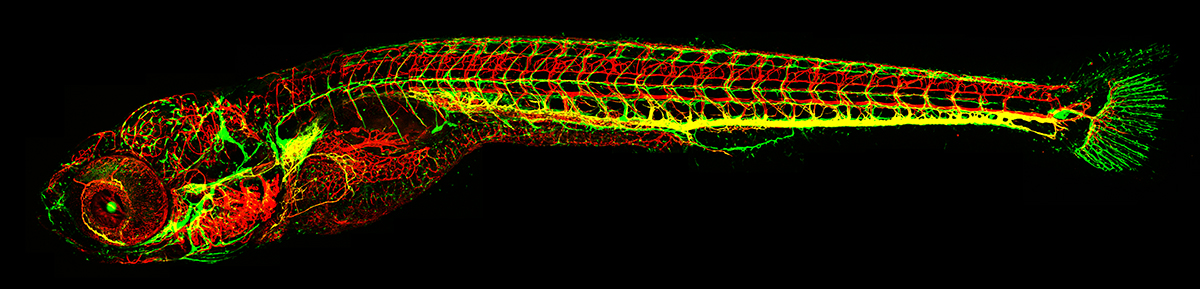
Hyun Min Jung, PhD
Section on Vertebrate Organogenesis (Weinstein Lab)
This is a 13-day old transgenic zebrafish labeling blood vessels with red fluorescence protein and lymphatic vessels with green fluorescence protein. The mCherry and GFP expression is driven by kdrl and mrc1 gene promoter, respectively. This transgenic animal is a powerful tool to investigate the formation of complex vasculature networks during embryonic development in real-time.
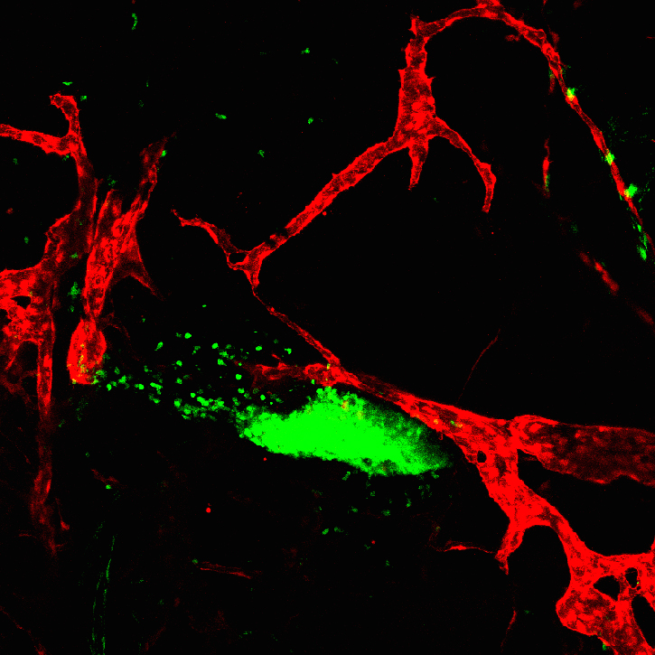
Hyun Min Jung, PhD
Section on Vertebrate Organogenesis (Weinstein Lab)
Migration of lymphocytes from thymus (GFP labeled oval shape structure) toward lymphatic vessels (labeled with RFP). Lck promoter drives GFP expression and lyve1 promoter drives RFP expression.
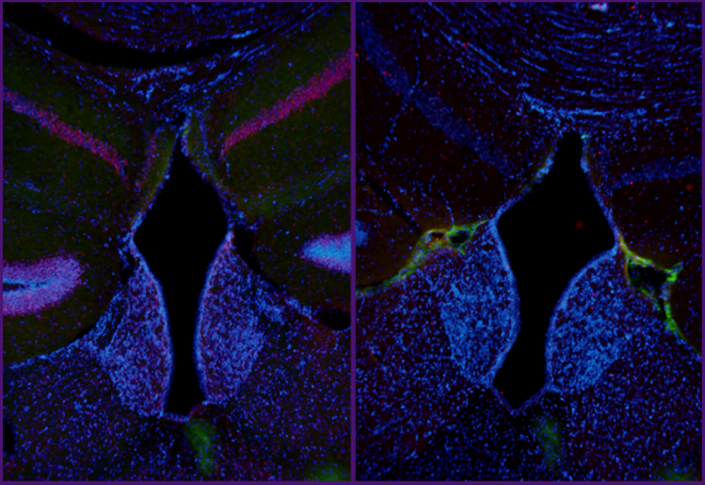
Edra London, PhD
Section on Endocrinology and Genetics (Stratakis Lab)
Resubmission from 2016
Brain sections from a Prkar2a knockout (left) and wild type (right) mouse were subjected to immunofluorescent staining following two weeks of free access to running wheels followed by blocking of running activity by locking the running wheels. Sections were stained for beta actin (green), c-fos (red) and counterstained with DAPI. C-fos appears to be highly induced in dentate gyrus as well as medial and lateral habenula, brain regions associated with voluntary motor activity and reward. Prkar2a knockout mice resist diet-induced obesity and run more than wild-type mice when given access to running wheels.
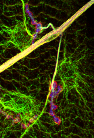
Qi Wang, PhD
Unit on Cellular Communication (Serpe Lab)
Resubmission from 2016
Cytoskeleton system at Drosophila NMJ. Green, microtuble cytoskeleton; Red, actin cytoskeleton; Blue, motor neuron surface.
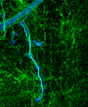
Qi Wang, PhD
Unit on Cellular Communication (Serpe Lab)
Resubmission from 2016
Cytoskeleton system at Drosophila NMJ. Green, microtuble cytoskeleton; Blue, motor neuron surface.
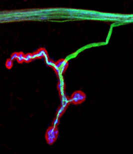
Qi Wang, PhD
Unit on Cellular Communication (Serpe Lab)
Resubmission from 2016
Cytoskeleton system at Drosophila NMJ. Green, microtuble cytoskeleton; Red, actin cytoskeleton; Blue, motor neuron surface.