
Past Retreats: 2016 Image Competition Entries
Click to view larger images.
The Committee also considered 2015 submissions from Matthew Butler, Sarah Cohen and Alex Valm, and Justin Demmerle.

Competition Winner:
Sarah Cohen, PhD, and Alex Valm, PhD
Section on Organelle Biology (Lippincott-Schwartz Lab)
Resubmission from 2015
Multicolor organelles in an African Green Monkey kidney cell. Purple, Endoplasmic reticulum; green, mitochondria; yellow, Golgi apparatus; cyan, lysosomes; red, peroxisomes; white, lipid droplets.
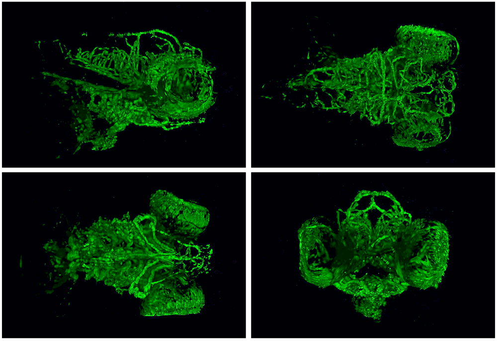
Matther Butler, PhD
Section on Vertebrate Organogenesis (Weinstein Lab)
Resubmission from 2015
High resolution SPIM images taken of 5 day old zebrafish heads. Images show blood vessels developing within the brain and eye of the fish.
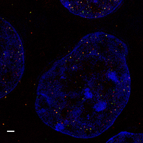
Justin Demmerle, BA
Unit on Mammalian Epigenome Reprogramming (Macfarlan Lab)
Resubmission from 2015
Super-resolution structured illumination microscopy (SIM) image of two cooperating transcription factors, red and green, within the nuclei (blue) of mouse embryonic stem cells. Scale bar = 1 micron.
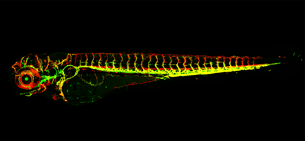
Hyun Min Jung, PhD
Section on Vertebrate Organogenesis (Weinstein Lab)
Confocal microscopy image of a 3-day old LGBVR (Lymphatic Green Blood Vessel Red) double transgenic zebrafish. Lymphatics and blood vasculatures are labeled with GFP and RFP, respectively.
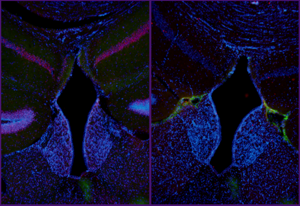
Edra London, PhD
Section on Endocrinology and Genetics (Stratakis Lab)
Brain sections from a Prkar2a knockout (left) and wild type (right) mouse were subjected to immunofluorescent staining following two weeks of free access to running wheels followed by blocking of running activity by locking the running wheels. Sections were stained for beta actin (green), c-fos (red) and counterstained with DAPI. C-fos appears to be highly induced in dentate gyrus as well as medial and lateral habenula, brain regions associated with voluntary motor activity and reward. Prkar2a knockout mice resist diet-induced obesity and run more than wild-type mice when given access to running wheels.
Diego Martinelli, MD, PhD
Section on Translational Neuroscience (Kaler Lab)
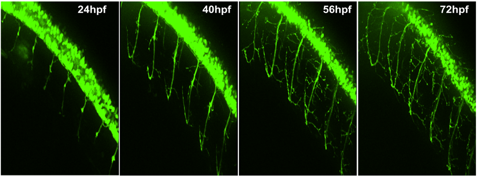
Hb9:GFP positive zebrafish allow in vivo motor neuron visualization.
Adult transgenic (hb9:GFP) zebrafish (Danio rerio, AB strain) express green fluorescent protein (GFP) driven by motor neuron-specific promoter Hb9. Time-lapse confocal imaging from 24 to 72 hours of motor neuron development in vivo in hHb9:GFP positive fish was performed with a Nikon Eclipse Spinning Disk equipped with a Perfect Focus attachment and a full stage-top incubator (CO2, temperature and humidity) and a CFI PlanApo VC 20x 0.75 NA air objective.
Alex Szatmary, PhD
Section on Cell Biophysics (Nossal Lab)
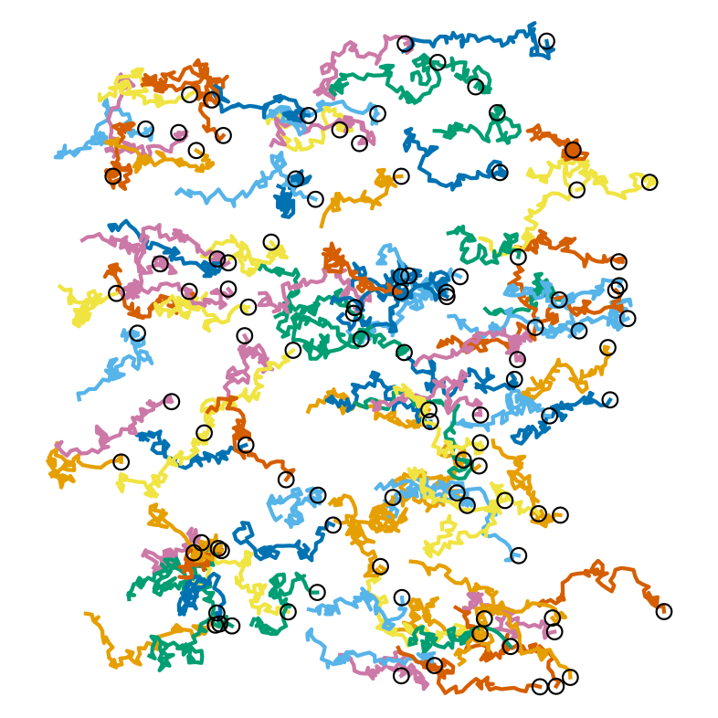
Neutrophil motion up a chemoattractant gradient is coordinated by gradient relay in these model results. Each line corresponds to the path taken by a single neutrophil over the course of an hour; circles indicate final positions. A primary chemoattractant gradient guides cells to the right, but the signal from this gradient can only be sensed reliably at the right half of this image. The cells on the right release vesicles that secrete a secondary gradient; this secondary gradient guides the cells on the left.
Giampaolo Trivellin, PhD
Section on Endocrinology and Genetics (Stratakis Lab)
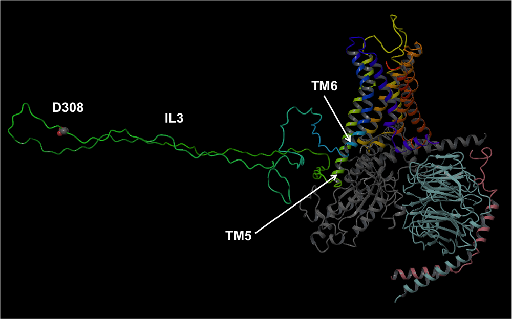
Predicted GPR101 3D structure in its activated state
Homology model of the human GPR101 in complex with a Gs heterotrimer. The p.E308D mutation is on the cytosolic side of the receptor, located in the long third intracellular loop (IL3), which connects transmembrane domains 5 and 6 (TM5 and TM6). The backbone of the receptor and the G protein heterotrimer is schematically represented as a ribbon, with the receptor shown with a spectrum of colors that ranges from red at the N-terminus to purple at the C-terminus, the alpha, beta and gamma subunits of the G protein are in gray, cyan and pink, respectively. The predicted GPR101 structure was determined in collaboration with Dr. Stefano Costanzi at American University.
Qi Wang, PhD
Unit on Cellular Communication (Serpe Lab)
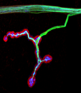
Cytoskeleton system at Drosophila NMJ. Green, microtuble cytoskeleton; Red, actin cytoskeleton; Blue, motor neuron surface.

Qi Wang, PhD
Unit on Cellular Communication (Serpe Lab)
Cytoskeleton system at Drosophila NMJ. Green, microtuble cytoskeleton; Blue, motor neuron surface.

Qi Wang, PhD
Unit on Cellular Communication (Serpe Lab)
Cytoskeleton system at Drosophila NMJ. Green, microtuble cytoskeleton; Red, actin cytoskeleton; Blue, motor neuron surface.