
Past Retreats: 2015 Image Competition Entries
Click to view larger images.
The Committee also considered Mik Sulkowski's submissions from 2013.
Competition Winner #1:
Mikolaj Sulkowski, PhD
Unit on Cellular Communication (Mihaela Serpe Lab)
3D structured illumination microscopy of the Drosophila neuromuscular junction reveals sub-synaptic localization of signaling molecules.
Competition Winner #2:
Mikolaj Sulkowski, PhD
Unit on Cellular Communication (Mihaela Serpe Lab)
3D structured illumination microscopy of the Drosophila neuromuscular junction reveals sub-synaptic localization of signaling molecules.
Competition Winner #3:
Mikolaj Sulkowski, PhD
Unit on Cellular Communication (Mihaela Serpe Lab)
3D structured illumination microscopy of the Drosophila neuromuscular junction reveals sub-synaptic localization of signaling molecules.
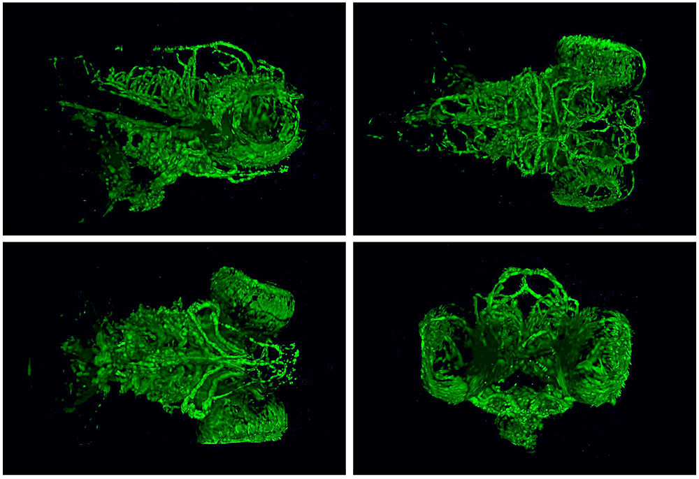
Matther Butler, PhD
Section on Vertebrate Organogenesis (Brant Weinstein Lab)
High resolution SPIM images taken of 5 day old zebrafish heads. Images show blood vessels developing within the brain and eye of the fish.

Sarah Cohen, PhD
Section on Organelle Biology (Jennifer Lippincott-Schwartz Lab)
Multicolor organelles in an African Green Monkey kidney cell. Purple, Endoplasmic reticulum; green, mitochondria; yellow, Golgi apparatus; cyan, lysosomes; red, peroxisomes; white, lipid droplets.

Justin Demmerle, BA
Unit on Mammalian Epigenome Reprogramming (Todd Macfarlan Lab)
Super-resolution structured illumination microscopy (SIM) image of two cooperating transcription factors, red and green, within the nuclei (blue) of mouse embryonic stem cells. Scale bar = 1 micron.

Amanda Dettmer, PhD
Comparative Behavioral Genetics Section (Steve Suomi Lab)
Researchers at the Laboratory of Comparative Ethology study the predictors of chronic stress, and subsequent influences on behavior, across the lifespan in semi-free ranging rhesus monkeys. Here, Dr. Amanda M. Dettmer (Postdoctoral IRTA) is being “escorted” in her data collection by the troop’s oldest member, Babette. Photo credit: Emma Soneson.

Annika Paukner, PhD
Comparative Behavioral Genetics Section (Steve Suomi Lab)
A rhesus monkey mother, Xadie, intensely focuses on her 6-week old infant, Eunice (named after NICHD’s benefactor). Researchers at the Laboratory of Comparative Ethology are studying the developmental consequences of mother-infant face-to-face interactions in semi-free ranging monkeys. Photo credit: Amanda Dettmer
Ian Williams, PhD
Section on Molecular Dysmorphology (Forbes Porter Lab)
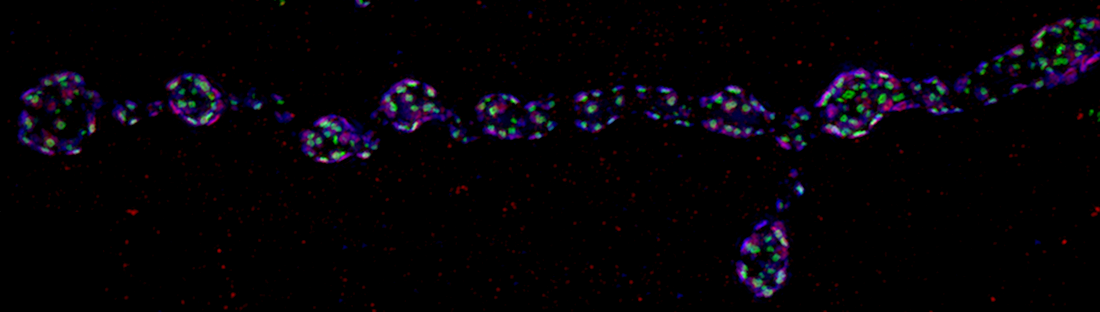
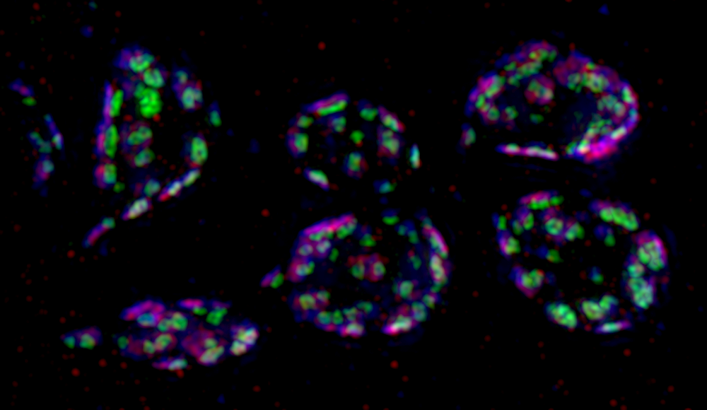
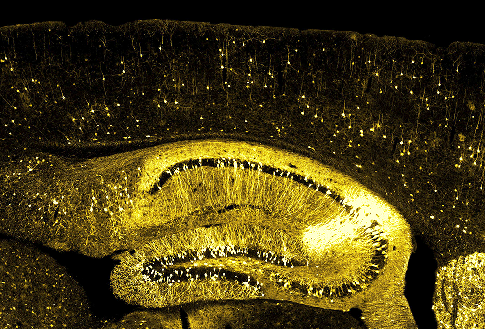
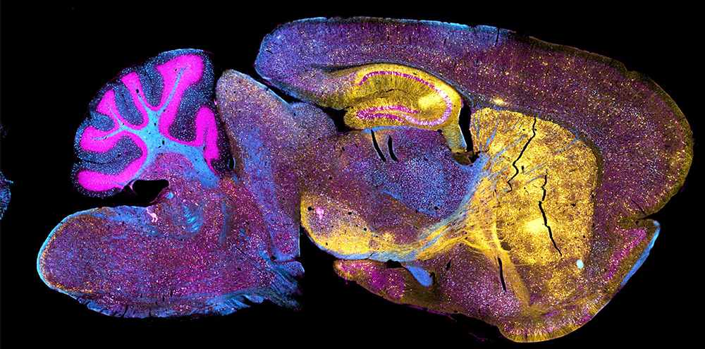
hippocampus yellow: Fluorescence labelled pictures of mouse brain from our NPC1 colony, the mouse model of the neurodegenerative disease called Niemann Pick type C1 we use in the lab. One major way of assessing the impact of a potential therapeutic in the CNS is to assess cerebellar pathology in these mice, namely the number of surviving Purkinje cell neurons, the condition of their long distance projection axons and their connectivity with their target neurons.
In NPC1 disease patients cognitive impairment is also a major symptom, but the forebrain in the NPC1 mouse model is not well-studied in comparison to the cerebellum. The various "GFP in brain" pictures show an NPC1 disease brain labelled to highlight forebrain structures, including hippocampal, striatal and cortical neurons (in green or yellow). The close ups in the remaining images show the cellular detail of these neurons nicely, including the single channel gold-colored hippocampus image in "hippocampus yellow", where just a simple stain can show the most detail.
Ian Williams, PhD
Section on Molecular Dysmorphology (Forbes Porter Lab)
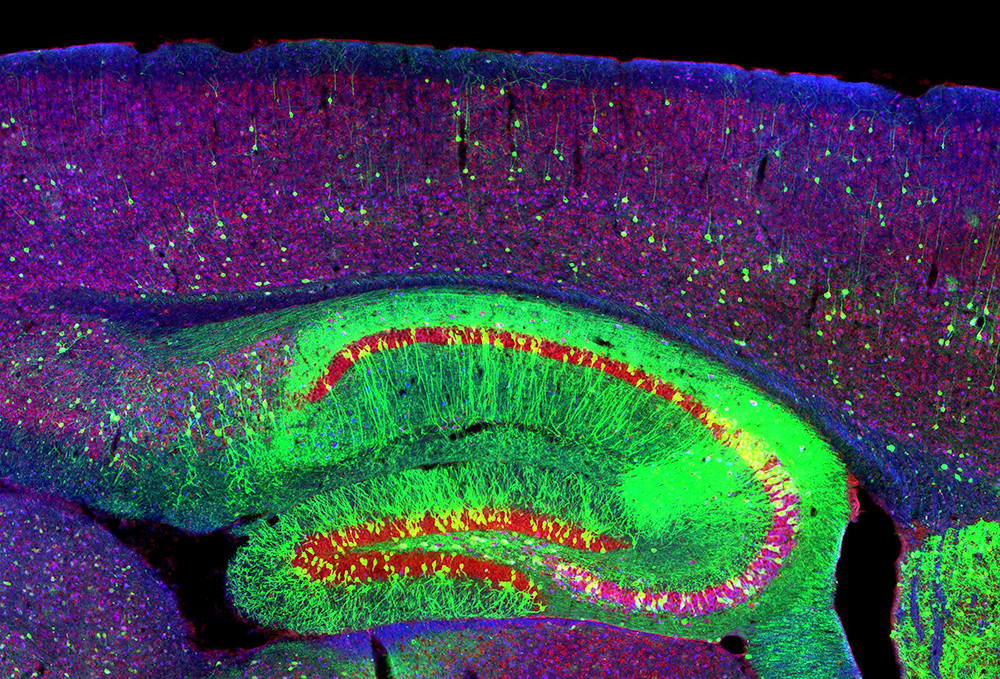
hippocampus rgb: Fluorescence labelled pictures of mouse brain from our NPC1 colony, the mouse model of the neurodegenerative disease called Niemann Pick type C1 we use in the lab. One major way of assessing the impact of a potential therapeutic in the CNS is to assess cerebellar pathology in these mice, namely the number of surviving Purkinje cell neurons, the condition of their long distance projection axons and their connectivity with their target neurons.
In NPC1 disease patients cognitive impairment is also a major symptom, but the forebrain in the NPC1 mouse model is not well-studied in comparison to the cerebellum. The various "GFP in brain" pictures show an NPC1 disease brain labelled to highlight forebrain structures, including hippocampal, striatal and cortical neurons (in green or yellow). The close ups in the remaining images show the cellular detail of these neurons nicely, including the single channel gold-colored hippocampus image in "hippocampus yellow", where just a simple stain can show the most detail.
Ian Williams, PhD
Section on Molecular Dysmorphology (Forbes Porter Lab)
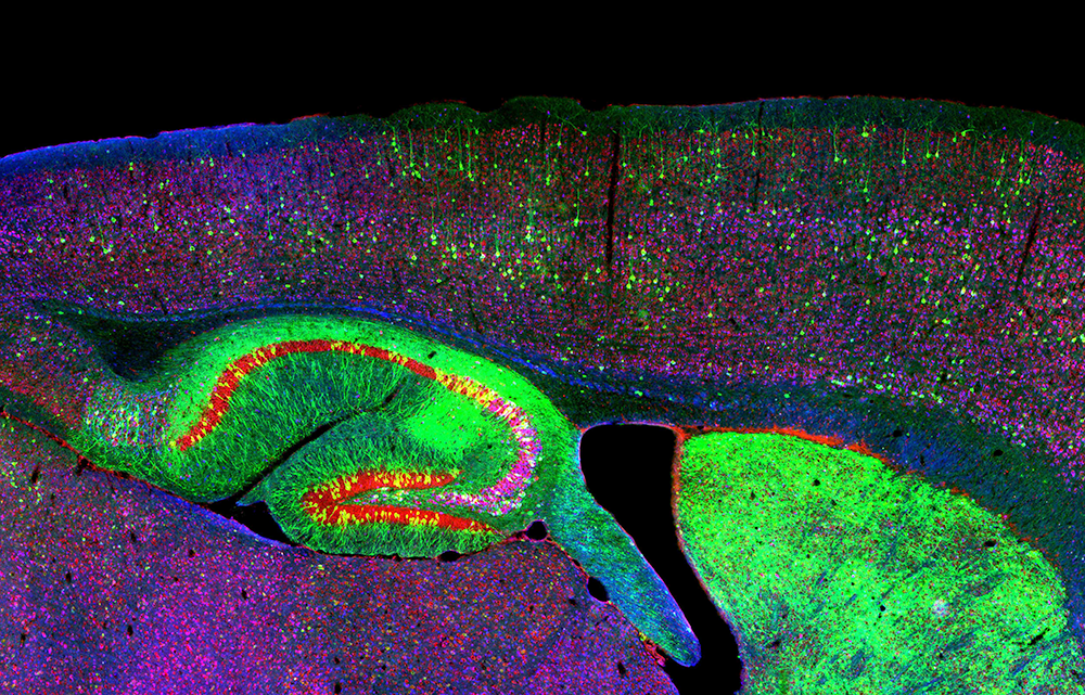
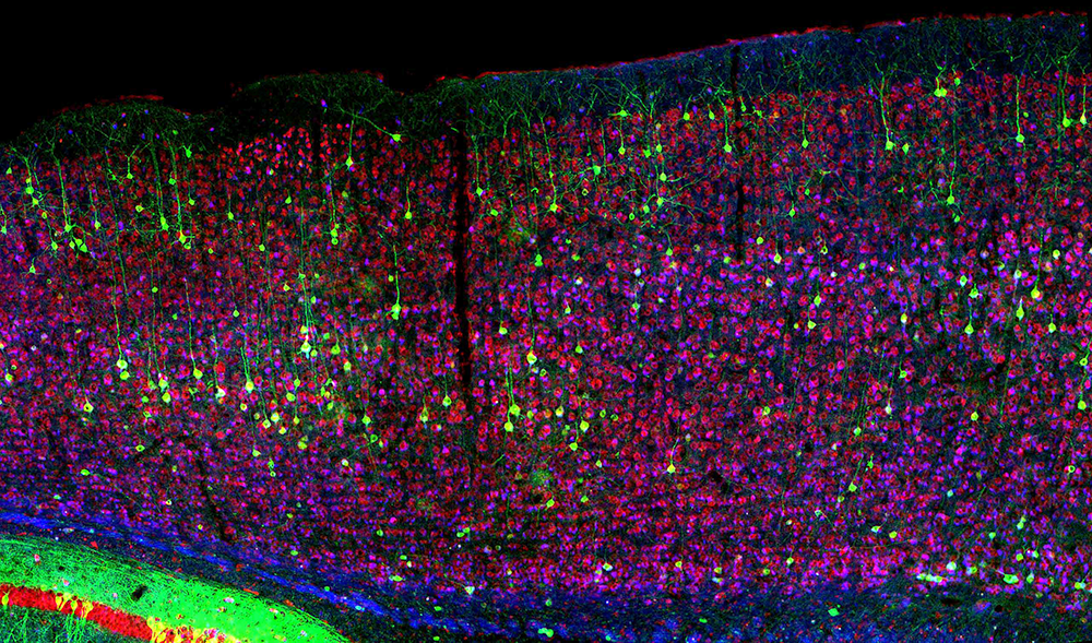
hip and cortex rgb: Fluorescence labelled pictures of mouse brain from our NPC1 colony, the mouse model of the neurodegenerative disease called Niemann Pick type C1 we use in the lab. One major way of assessing the impact of a potential therapeutic in the CNS is to assess cerebellar pathology in these mice, namely the number of surviving Purkinje cell neurons, the condition of their long distance projection axons and their connectivity with their target neurons.

Ian Williams, PhD
Section on Molecular Dysmorphology (Forbes Porter Lab)
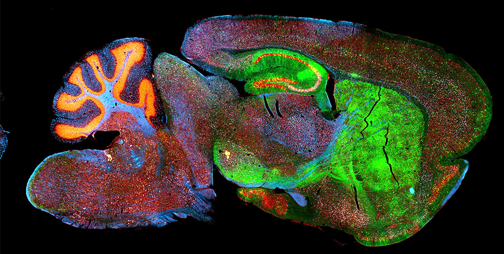
GFP in brain alt 1-1: Fluorescence labelled pictures of mouse brain from our NPC1 colony, the mouse model of the neurodegenerative disease called Niemann Pick type C1 we use in the lab. One major way of assessing the impact of a potential therapeutic in the CNS is to assess cerebellar pathology in these mice, namely the number of surviving Purkinje cell neurons, the condition of their long distance projection axons and their connectivity with their target neurons.
In NPC1 disease patients cognitive impairment is also a major symptom, but the forebrain in the NPC1 mouse model is not well-studied in comparison to the cerebellum. The various "GFP in brain" pictures show an NPC1 disease brain labelled to highlight forebrain structures, including hippocampal, striatal and cortical neurons (in green or yellow). The close ups in the remaining images show the cellular detail of these neurons nicely, including the single channel gold-colored hippocampus image in "hippocampus yellow", where just a simple stain can show the most detail.
Ian Williams, PhD
Section on Molecular Dysmorphology (Forbes Porter Lab)
GFP in brain alt 2-1: Fluorescence labelled pictures of mouse brain from our NPC1 colony, the mouse model of the neurodegenerative disease called Niemann Pick type C1 we use in the lab. One major way of assessing the impact of a potential therapeutic in the CNS is to assess cerebellar pathology in these mice, namely the number of surviving Purkinje cell neurons, the condition of their long distance projection axons and their connectivity with their target neurons.
In NPC1 disease patients cognitive impairment is also a major symptom, but the forebrain in the NPC1 mouse model is not well-studied in comparison to the cerebellum. The various "GFP in brain" pictures show an NPC1 disease brain labelled to highlight forebrain structures, including hippocampal, striatal and cortical neurons (in green or yellow). The close ups in the remaining images show the cellular detail of these neurons nicely, including the single channel gold-colored hippocampus image in "hippocampus yellow", where just a simple stain can show the most detail.
Ian Williams, PhD
Section on Molecular Dysmorphology (Forbes Porter Lab)
GFP in brain alt 3-1: Fluorescence labelled pictures of mouse brain from our NPC1 colony, the mouse model of the neurodegenerative disease called Niemann Pick type C1 we use in the lab. One major way of assessing the impact of a potential therapeutic in the CNS is to assess cerebellar pathology in these mice, namely the number of surviving Purkinje cell neurons, the condition of their long distance projection axons and their connectivity with their target neurons.
In NPC1 disease patients cognitive impairment is also a major symptom, but the forebrain in the NPC1 mouse model is not well-studied in comparison to the cerebellum. The various "GFP in brain" pictures show an NPC1 disease brain labelled to highlight forebrain structures, including hippocampal, striatal and cortical neurons (in green or yellow). The close ups in the remaining images show the cellular detail of these neurons nicely, including the single channel gold-colored hippocampus image in "hippocampus yellow", where just a simple stain can show the most detail.
Ian Williams, PhD
Section on Molecular Dysmorphology (Forbes Porter Lab)
cortex rgb: Fluorescence labelled pictures of mouse brain from our NPC1 colony, the mouse model of the neurodegenerative disease called Niemann Pick type C1 we use in the lab. One major way of assessing the impact of a potential therapeutic in the CNS is to assess cerebellar pathology in these mice, namely the number of surviving Purkinje cell neurons, the condition of their long distance projection axons and their connectivity with their target neurons.

Ian Williams, PhD
Section on Molecular Dysmorphology (Forbes Porter Lab)
++ npc cereb pop art: Fluorescence labelled pictures of mouse brain from our NPC1 colony, the mouse model of the neurodegenerative disease called Niemann Pick type C1 we use in the lab. One major way of assessing the impact of a potential therapeutic in the CNS is to assess cerebellar pathology in these mice, namely the number of surviving Purkinje cell neurons, the condition of their long distance projection axons and their connectivity with their target neurons.
In "++ cereb pop art", a cerebellar section has been stained for Purkinje cells, the white matter tracts with filipin (a label of unesterified cholesterol), and the other neurons with a fluorescent nissl stain. The three panels are exactly the same image with the same underlying data, just pseudocoloured differently. Different color combinations can provide better contrast for the eye for different cellular details, the standard rgb gives good general image, but using cyan magenta yellow in different orders can help pick out double labeling more easily (the blue and yellow combining in the centre panel to pick out the double labeling of Purkinje cells in green), or the yellow axons contrasting against the blue myelin to see axonal structure more easily in the right panel.

Ian Williams, PhD
Section on Molecular Dysmorphology (Forbes Porter Lab)
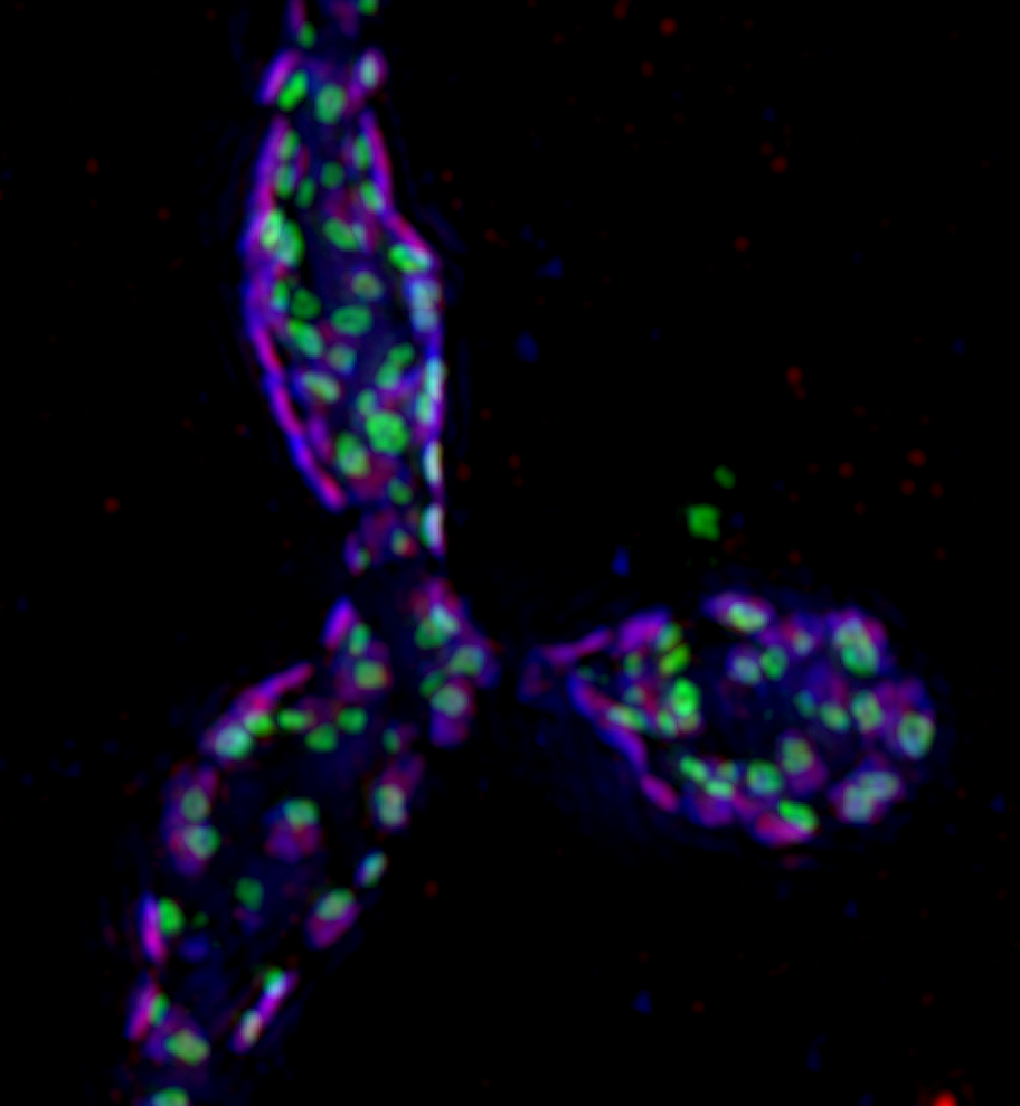
++ npc cereb butterfly: Fluorescence labelled pictures of mouse brain from our NPC1 colony, the mouse model of the neurodegenerative disease called Niemann Pick type C1 we use in the lab. One major way of assessing the impact of a potential therapeutic in the CNS is to assess cerebellar pathology in these mice, namely the number of surviving Purkinje cell neurons, the condition of their long distance projection axons and their connectivity with their target neurons.
"++ npc cereb butterfly" is the same image, just mirrored and aligned for aesthetic effect.
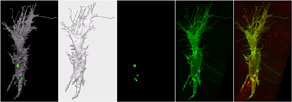
Alex Yu, PhD
Section on Vertebrate Organogenesis (Brant Weinstein Lab)
A single endothelial cell (green fluorescence) within the developing blood vessel (red fluorescence) was captured from the living zebrafish at 28 hour post fertilization. The fine membrane structures including filopodia and intracellular vesicles are highlighted in the surface-rendering images (grey).