
Past Retreats: 2011 Image Competition Entries
Click to view larger images.
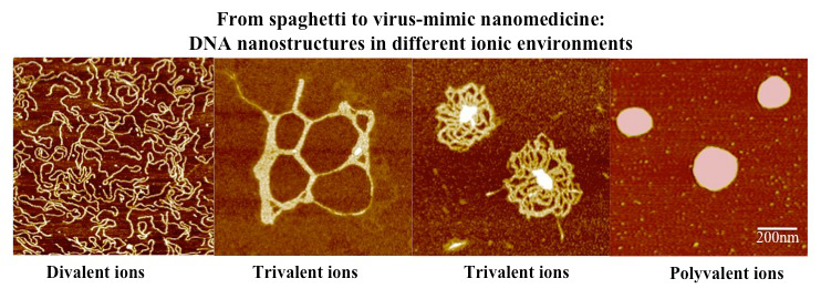
Competition Winner:
Preethi Chandran, PhD
Section on Tissue Biophysics and Biomimetics (Peter Basser Lab)
Atomic Force Imaging shows that DNA forms complex nanostructures with multivalent ions. The interaction can be modulated to obtain virus-like particles which serve as nanomedicines that deliver a anti-retroviral genes to compromised cells. Each image area is 1um X 1um.
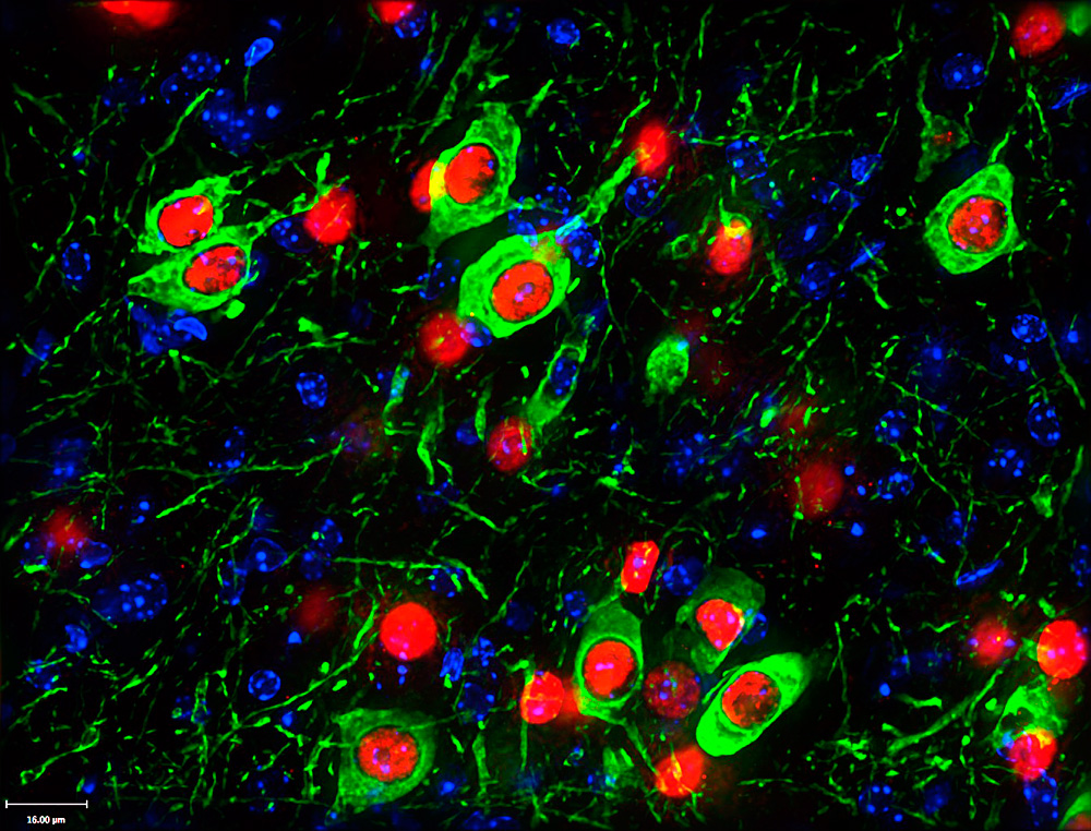
Lindsay Hayes, PhD
Unit on Developmental Neurogenetics (Sohyun Ahn Lab)
The Sonic Hedgehog lineage (red) greatly contributes to the midbrain dopamine neurons (green), however some cells develop independent of the Sonic Hedgehog lineage (green without red).
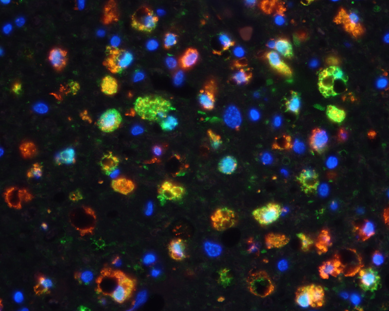
Suh Young Jeong, PhD
Section on Human Iron Metabolism (Tracey Rouault Lab)
Lumbar spinal cord section from an ALS (Amyotrophic Lateral Sclerosis) patient stained with anti-CD68 (red, macrophage) and anti-ferritin (green, iron storage protein) antibodies. DAPI (blue) was used to visualize nucleus.
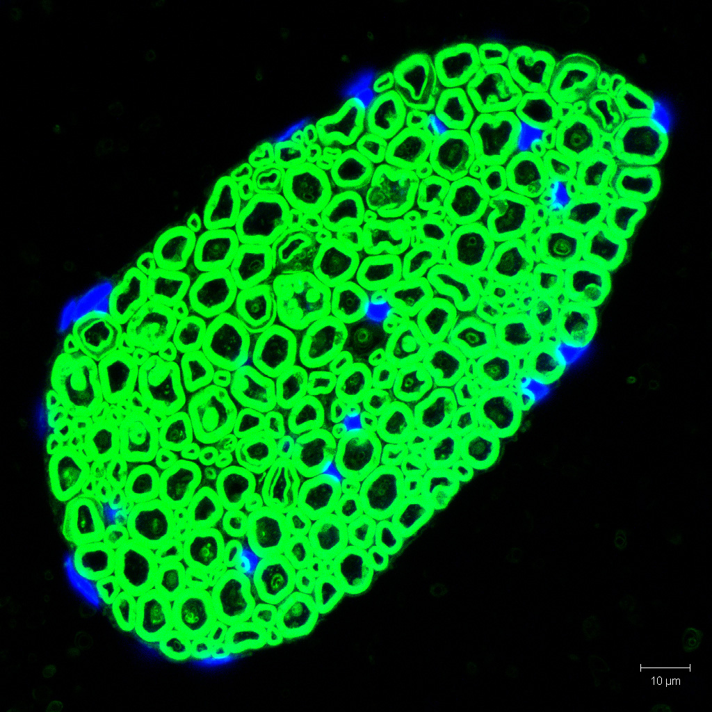
Suh-Young Jeong, PhD
Section on Human Iron Metabolism (Tracey Rouault Lab)
Mouse ventral root nerve section stained with BODIPY lipid probe (green). Each green ring represent a myelinated axon and DAPI (blue) was used to counterstain nucleus.
Mikolaj Sulkowski, PhD
Unit on Cellular Communication (Mihaela Serpe Lab)
Confocal image of a Drosophila ventral ganglion stained for pMad5 and Repo