
Past Retreats: 2009 Image Competition Entries
Click to view larger images.
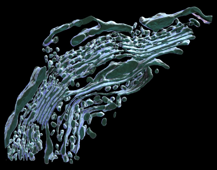
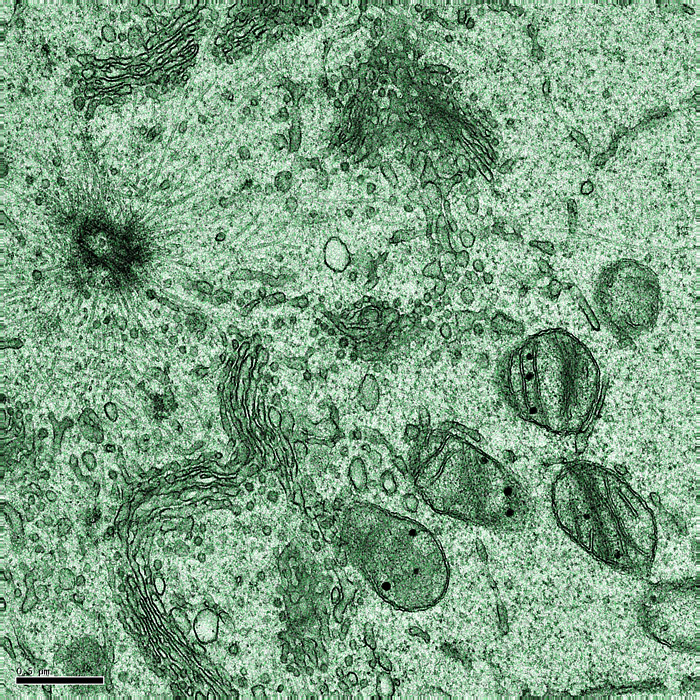
Competition Winner:
Rachid Sougrat, PhD
Section of Organelle Biology (Jennifer Lippincott-Schwartz Lab)
The picture is a 3D rendering of the most fascinating organelle in a mammalian cell: the Golgi Apparatus. This piece of Golgi ribbon, from a mouse spermatid, was imaged with an electron microscope using the tomography technique. Its complex architecture illustrates the complexity of its function.
Jun Chen, PhD
Section on Endocrine Physiology (Greti Aguilera Lab)
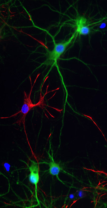
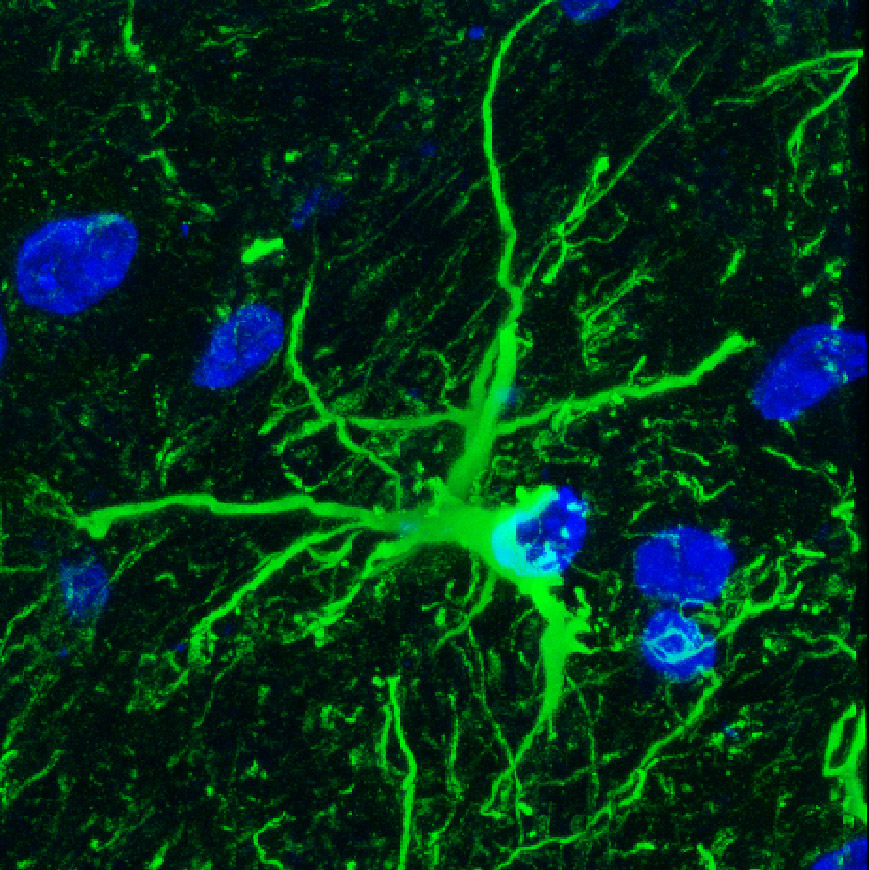
Neuron
Jun Chen, PhD
Section on Endocrine Physiology (Greti Aguilera Lab)
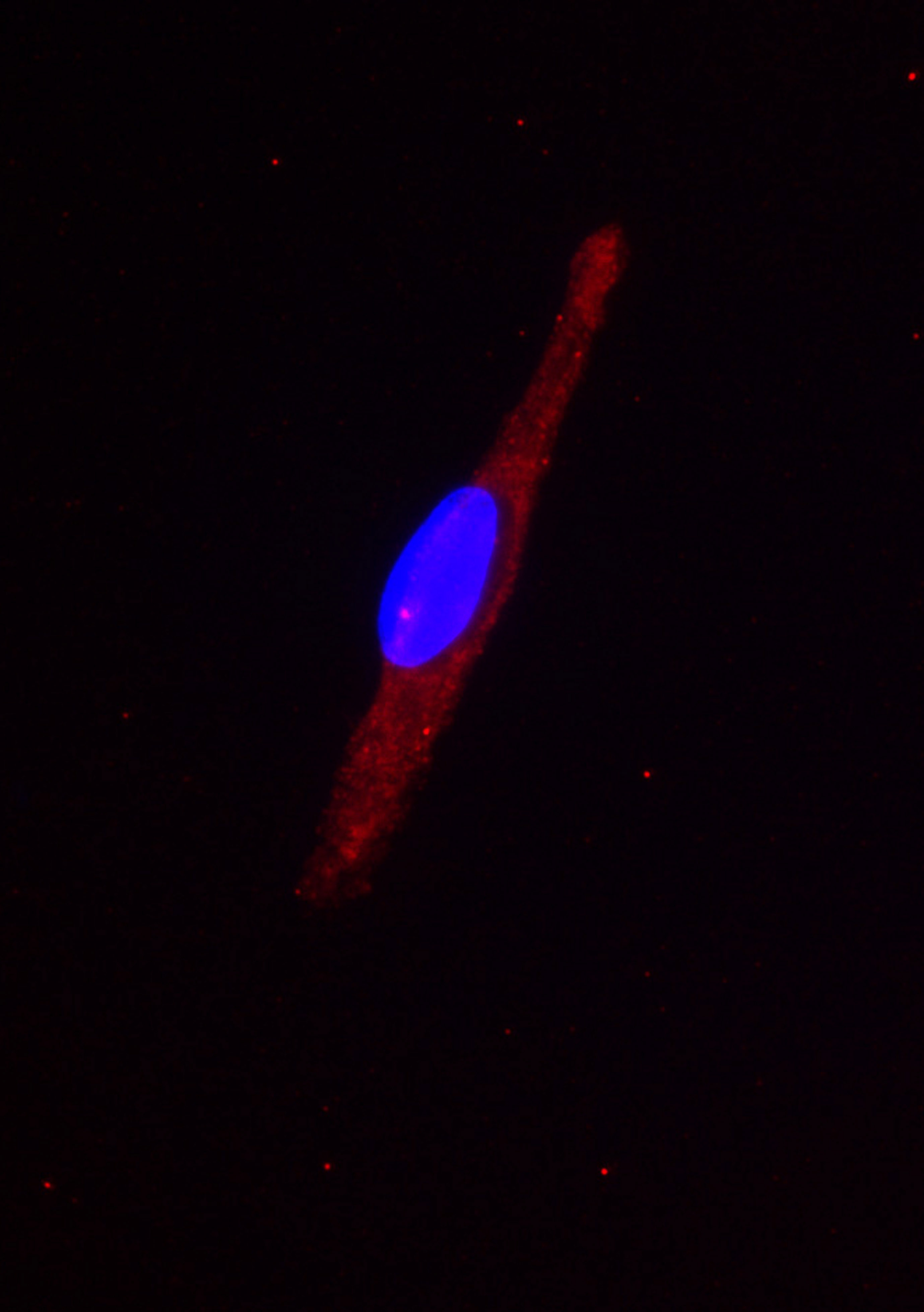
H32 Cells
Jun Chen, PhD
Section on Endocrine Physiology (Greti Aguilera Lab)
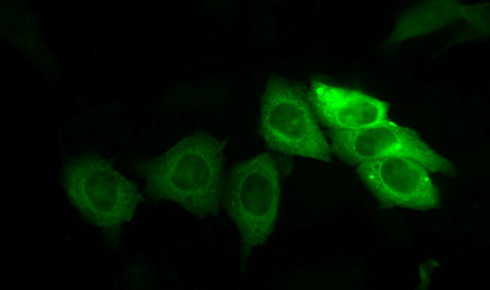
H32 GFP 3

Ariel Ginzberg, PhD
Section on Bacterial Disease Pathogenesis and Immunity (Rachel Schneerson Lab)
Dark Hollow Falls, Shenandoah National Park, Virginia
Suh Young Jeong, PhD
Section on Human Iron Metabolism (Tracey Rouault Lab)
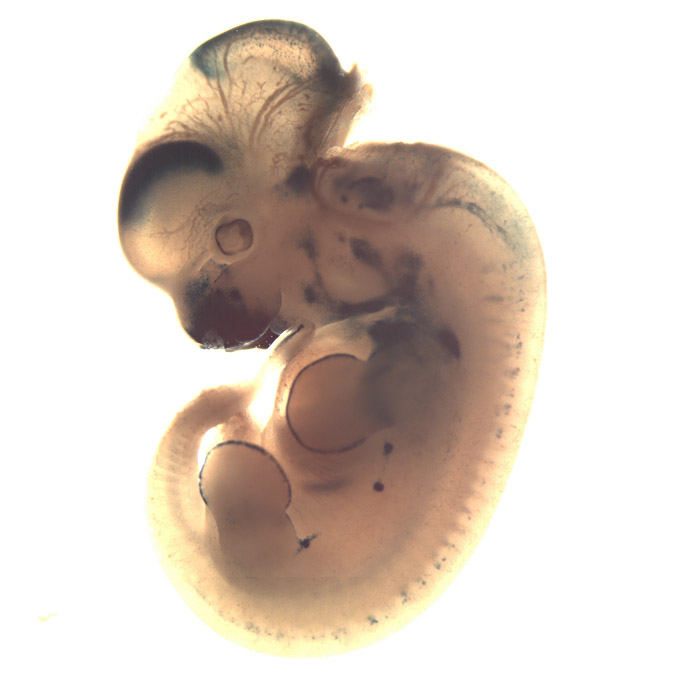
Matthew Phillips, PhD
Section on Mammalian Molecular Genetics (Heiner Westphal Lab)
Mouse embryo
Rachid Sougrat, PhD
Section of Organelle Biology (Jennifer Lippincott-Schwartz Lab)
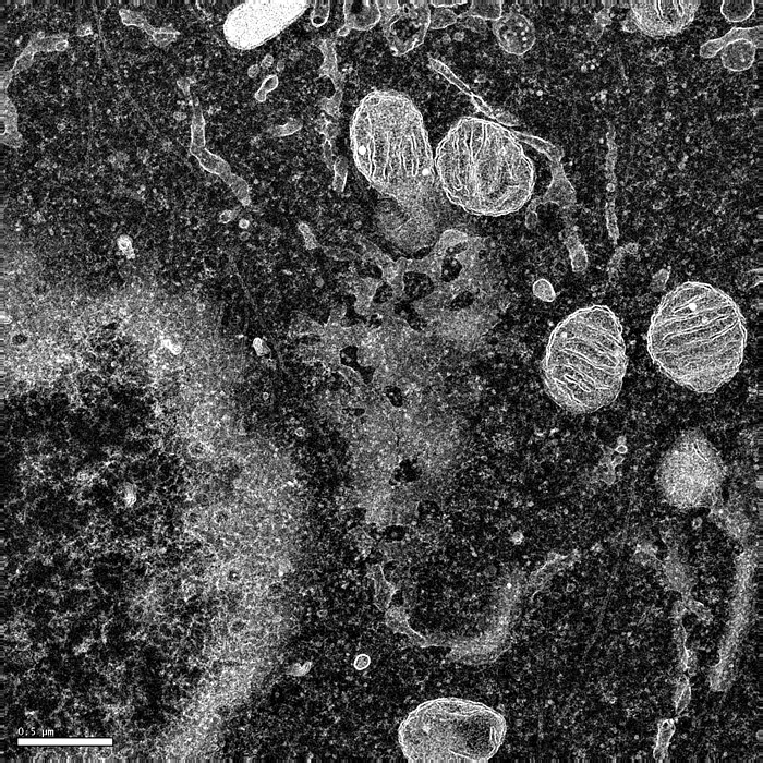
Rachid Sougrat, PhD
Section of Organelle Biology (Jennifer Lippincott-Schwartz Lab)
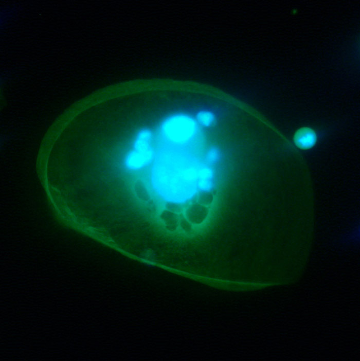
Wenge Zhu, PhD
Section on Eukaryotic Gene Regulation (Melvin L. DePamphilis Lab)
Two cancer cells (breast cancer cells). Both cells are GFP labeled. Big cell in center is a dying cell with re-replicated DNA and falling apart nucleus, induced by depletion of geminin, a DNA replication inhibitor. The giant cell just de-attached the culture dish. The small cells on the shoulder of giant cell is untreated cells with normal size. Light blue is nuclei and green color represents the whole cell.